Aptagen, LLC

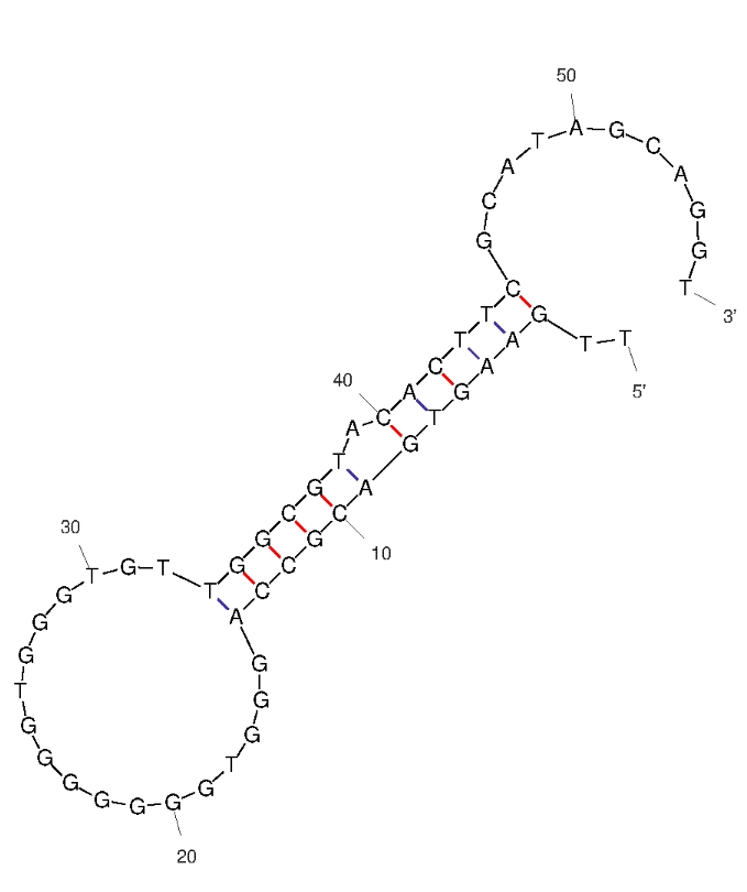
A24-3 (ID# 9049)

DNA
bPAG9
Protein
2.02±0.28 nM (reported value)
The truncated aptamer was diluted to 2 μM in binding bufer and mixed with an equal volume of bPAG9 (fnal concentration, 1.5 μM), with binding bufer used as a control. Each
CD spectrum was obtained by accumulating two scans at
100 nm min⁻1with an instrument time constant of 1 s.
Data were collected from 220 to 350 nm at 1 nm intervals. The background spectrum (binding bufer) was subtracted from the CD data.
25 °C°C
NA If the oligo is a known aptamer sequence: For binding studies, perform a refolding protocol to ensure proper function (i.e. binding to antigen or target). Refer to the aptamer reference source for the appropriate refolding parameters and binding conditions. Note: it is unknown whether aptamer functions properly without refolding.
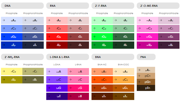
dTpdTpdGpdApdApdGpdTpdGpdApdCpdGpdCpdCpdApdGpdGpdGpdTpdGpdGpdGpdGpdGpdGpdGpdTpdGpdGpdGpdTpdGpdTpdTpdGpdGpdCpdGpdTpdApdCpdApdCpdTpdTpdCpdGpdCpdApdTpdApdGpdCpdApdGpdGpdTp


56
17544.3 g/mole
541400 L/(mole·cm)
Note: Information on this aptamer oligo was obtained from the literature and hasn't been validated by Aptagen.
Lu, C., Qin, J., Wu, S. et al. Structural optimization, characterization, and evaluation of binding mechanism of aptamers against bovine pregnancy-associated glycoproteins and their application in establishment of a colorimetric aptasensor using Fe-based metal-organic framework as peroxidase mimic tags. Microchim Acta 191, 713 (2024).
Have your aptamer oligo synthesized ORDER


We are always looking for ways to improve. Please tell us what you think.